
Written by a Caring Mom at Parenting Stories
Fetal heart development starts within the first weeks after conception. At first, the heart is just a straight tube. Over time, it curves and forms loops. Then it slowly turns into a structure with chambers and valves. It also begins to connect with the large blood vessels. By the end of the first trimester, the main layout is mostly there. Even so, the heart still keeps growing and refining its parts all the way up to birth.
For many expectant parents, that small moving spot on an early ultrasound can make the pregnancy feel real right away. It can also trigger a lot of questions. When do you hear or see the heartbeat for the first time? What changes during the 12-week mark? What will the later anatomy scan show about the baby’s heart?
In this guide, we will walk through how the heart develops during pregnancy. We will also cover what you might notice at key ultrasound check points.
When Does Fetal Heart Development Begin?
Your baby’s heart begins developing very early, during the first few weeks after conception. At this stage, your baby is still incredibly tiny, but important changes are already happening.
The heart doesn’t appear as a fully formed four-chambered organ right away. It begins as a simple heart tube made from specialized cells. As your baby grows, this tube lengthens, bends, and loops into a more complex shape.
Over the following weeks, the developing heart gradually forms chambers, valves, and major blood vessels that will help circulate blood throughout your baby’s growing body.
All of this starts before you can feel your baby move and often before you even have a noticeable pregnancy bump. It’s one of the earliest and most fascinating parts of your baby’s development.
When Does a Baby’s Heart Start Beating?
Rhythmic cardiac activity begins early, while the heart is still developing.
Around 5 to 6 weeks of pregnancy, this activity may sometimes be visible on a transvaginal ultrasound. At this stage, however, the heart isn’t yet a fully developed four-chambered heart. Cleveland Clinic explains that the cells that will form the heart begin clustering and pulsing around this stage of pregnancy.
This is also why very early ultrasound results need to be interpreted carefully.
If cardiac activity isn’t visible during an early scan, it doesn’t automatically mean something is wrong. Your dates may be slightly off, especially if you ovulated later than expected.
Your healthcare provider may recommend another ultrasound after an appropriate interval.
Fetal Heart Development Week by Week
The biggest structural changes happen during the early weeks of pregnancy.
Around Weeks 5–6
The early heart tube continues developing and changing shape. Cardiac activity may become visible on ultrasound around this stage, depending on the actual gestational age and type of scan.
For many parents, seeing that tiny flicker is the first moment pregnancy feels truly real. But don’t compare your scan with someone else’s. A difference of just a few days can affect what is visible this early.
Around Weeks 6–8
The developing heart becomes much more complex. The heart tube bends and loops, while structures that contribute to the chambers and internal walls continue developing. Major blood vessels connected to the heart are also taking shape. MedlinePlus notes that during weeks 6 to 7, the heart continues growing, develops a regular rhythm, and blood begins pumping through the main vessels.
The heart will continue to grow throughout pregnancy, but these early weeks establish much of its basic structure.
When Does Fetal Pole Develop?
The fetal pole usually becomes visible around 5½ to 6 weeks of pregnancy on a transvaginal ultrasound, although the exact timing can vary. It is an early ultrasound term for the developing embryo and is often one of the first signs of your growing baby that can be seen.
If you’re around six weeks and the fetal pole isn’t visible yet, it doesn’t always mean something is wrong. Pregnancy dating is based on the first day of your last menstrual period, so later ovulation or conception can mean your pregnancy is actually a few days younger than expected.
When an ultrasound is simply too early to give a clear answer, your healthcare provider may recommend a repeat scan after an appropriate interval to check for further development and cardiac activity.
What Happens During 12 Week Fetal Development?
By 12 weeks, your baby has developed many of the basic body structures and is now entering a stage of continued growth and maturation. During 12 week fetal development, the fingers and toes are formed, facial features are becoming more defined, and your baby can make small movements even though you won’t usually feel them yet.
The heart has also come a long way. Its basic four-chamber structure has developed, and it is actively pumping blood through your baby’s growing body. The heart will continue to grow and mature throughout the rest of pregnancy.
Around this time, your healthcare provider may be able to detect your baby’s cardiac activity with a handheld Doppler. If it isn’t heard right away, try not to worry. Your exact gestational age, your baby’s position, and other factors can affect how easily it can be detected this early.
How Does Your Baby Get Oxygen Before Birth?
Before birth, your baby gets oxygen from the placenta through the umbilical cord, not by breathing through the lungs. This is why fetal circulation works differently from the circulation your baby will have after birth.
The developing lungs aren’t yet responsible for taking oxygen from the air. Instead, oxygen-rich blood from the placenta travels to your baby, while special pathways help direct blood around the lungs.
Two important pathways make this possible. The foramen ovale allows blood to pass between the heart’s upper chambers, while the ductus arteriosus helps direct blood away from the lungs.
These structures are a normal part of fetal development. After birth, when your baby takes those first breaths and the lungs begin working, circulation changes and these temporary pathways are no longer needed in the same way.
What Happens to the Heart After Birth?
Your baby’s first breaths trigger major changes in circulation.
When the lungs expand, blood begins flowing through them differently. Pressure inside the heart changes, and the special pathways needed during fetal life are no longer required in the same way.
The baby’s circulation gradually transitions from relying on the placenta for oxygen to using the lungs.
It’s an incredible change that begins around the time you finally get to hold your baby in your arms.
Which Fetal Development Occurs During the Second Trimester?
This period includes rapid growth and continued organ development.
Between roughly weeks 14 and 27, your baby’s bones become stronger, hearing develops, movements become more noticeable, and the brain, lungs, muscles, and heart continue maturing.
This is also when many moms begin feeling their baby’s movements.
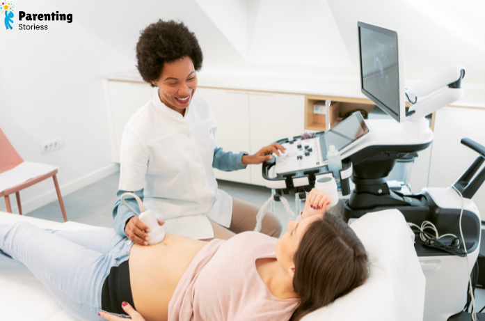
Another important milestone during this trimester is the detailed anatomy ultrasound.
What Is a Fetal Echocardiogram?
A fetal echocardiogram is a detailed ultrasound focused specifically on your baby’s heart.
It provides a closer look at the heart’s structure, rhythm, blood flow, and function.
When needed, a fetal echocardiogram is commonly performed around 18 to 22 weeks of pregnancy.
Not every pregnant woman needs one.
Your healthcare provider may recommend a fetal echo if the routine ultrasound shows something that needs a closer look or if certain family-history, maternal-health, pregnancy, or genetic factors increase the chance of a heart condition.
If you’re referred for one, try not to assume it means something is definitely wrong. Sometimes doctors simply need better or more detailed images.
What Are Congenital Heart Defects?
A congenital heart defect (CHD) is a problem with the heart’s structure or function that is present at birth.
Some congenital heart defects are mild, while others need specialized medical or surgical care. One example is tetralogy of Fallot, a congenital heart defect involving four problems with the heart and its blood vessels. According to the CDC, it may sometimes be detected during pregnancy through ultrasound, with a fetal echocardiogram used when a closer look at the baby’s heart is needed.
Congenital heart defects are among the most common types of birth defects. In many cases, there isn’t one clear reason why they happen.
Genetic factors can contribute to some heart conditions, while certain maternal health and environmental factors may also affect risk.
If a heart condition is detected during pregnancy, prenatal diagnosis can help doctors and parents prepare. Depending on the condition, specialists can plan where the baby should be delivered and what care may be needed after birth.
And if you’re facing this situation, don’t automatically blame yourself. A heart condition doesn’t necessarily mean something you did caused it.
How Does Fetal Lung Development Connect With the Heart?
Fetal lung development starts early and continues throughout pregnancy.
Before birth, however, your baby’s lungs aren’t responsible for getting oxygen into the bloodstream. The placenta handles that job.
As pregnancy progresses, the lungs grow and become more complex. Later, they begin producing surfactant, a substance that helps the tiny air spaces in the lungs stay open after birth.
Lung maturation continues during the third trimester.
Once your baby takes that first breath, the lungs begin providing oxygen and the heart’s circulation adjusts to work with them.
Can You Support Healthy Fetal Heart Development?
You can’t control every part of your baby’s development, but healthy pregnancy habits can support your overall health and your baby’s growth.
Keep your prenatal appointments and take prenatal vitamins or supplements as recommended by your healthcare provider.
Eat a balanced diet when you can, follow your provider’s advice about safe physical activity, and avoid alcohol, smoking, and recreational drugs.
Tell your doctor about prescription medicines, over-the-counter medicines, and herbal supplements you’re using. Don’t stop an important medication on your own—ask your healthcare provider what is safest during pregnancy.
If you have diabetes or another health condition, work with your healthcare team to manage it throughout pregnancy.
You don’t have to have a “perfect” pregnancy. Regular prenatal care and getting help when you need it matter much more than trying to do everything perfectly.
What If Cardiac Activity Isn’t Seen on an Early Ultrasound?
An early ultrasound without visible cardiac activity can be frightening, but timing matters.
Pregnancy dates aren’t always exact. If you ovulated later than expected, your pregnancy may be several days younger than the date calculated from your last period suggests.
Those few days can make a big difference during early pregnancy.
Your healthcare provider will consider your gestational age, ultrasound measurements, symptoms, and other clinical information before interpreting the results.
Sometimes another ultrasound after an appropriate interval is needed to understand how the pregnancy is developing.
Try not to compare your scan with ultrasound images online. They can’t tell you what’s happening in your individual pregnancy.
Helpful Questions to Ask at Your Ultrasound
It’s easy to forget your questions once you’re lying in the ultrasound room, so I like the simple idea of writing them down beforehand.
You can ask whether your baby’s growth matches the estimated gestational age, whether the heart rate and rhythm appear appropriate, whether the necessary heart views were obtained, and whether another ultrasound is needed.
If your provider recommends a fetal echocardiogram, ask why it’s needed and what they’re hoping to learn.
And if you don’t understand a medical term, ask them to explain it in simpler words. You deserve to understand what’s happening with your pregnancy.
Final Thoughts
Fetal heart development is an amazing process that starts in the earliest weeks of pregnancy. From a simple heart tube, your baby’s heart develops chambers, valves, and blood vessels and continues growing until birth.
Every pregnancy develops at its own pace, so early ultrasound findings may not always match an exact week or date. Keep up with your prenatal visits and ask your healthcare provider whenever you have questions about your baby’s heart or development.
From that first tiny flicker on an ultrasound to the heartbeat you hear at prenatal visits, each stage is another sign of how much your little baby is growing.
Want more simple, mom-friendly pregnancy guidance? Explore Parenting Stories for helpful tips and support through every stage of your pregnancy journey.
FAQs
1. When is the first ultrasound usually done?
The first ultrasound is often done between 6 and 12 weeks, depending on your pregnancy and your healthcare provider.
2. Can you see a baby at 4 weeks pregnant?
At 4 weeks, it may be too early to see much. Sometimes a small gestational sac is visible, but sometimes nothing clear appears yet.
3. When can a heartbeat be seen on ultrasound?
Cardiac activity may sometimes be visible around 6 weeks, although timing can vary.
4. Is a transvaginal ultrasound common in early pregnancy?
Yes. It is commonly used in early pregnancy because it can provide a clearer view than an abdominal scan.
Disclaimer
This article is for general educational purposes only and doesn’t replace professional medical advice, diagnosis, or prenatal care. Always speak with your healthcare provider about concerns regarding your pregnancy or your baby’s development.